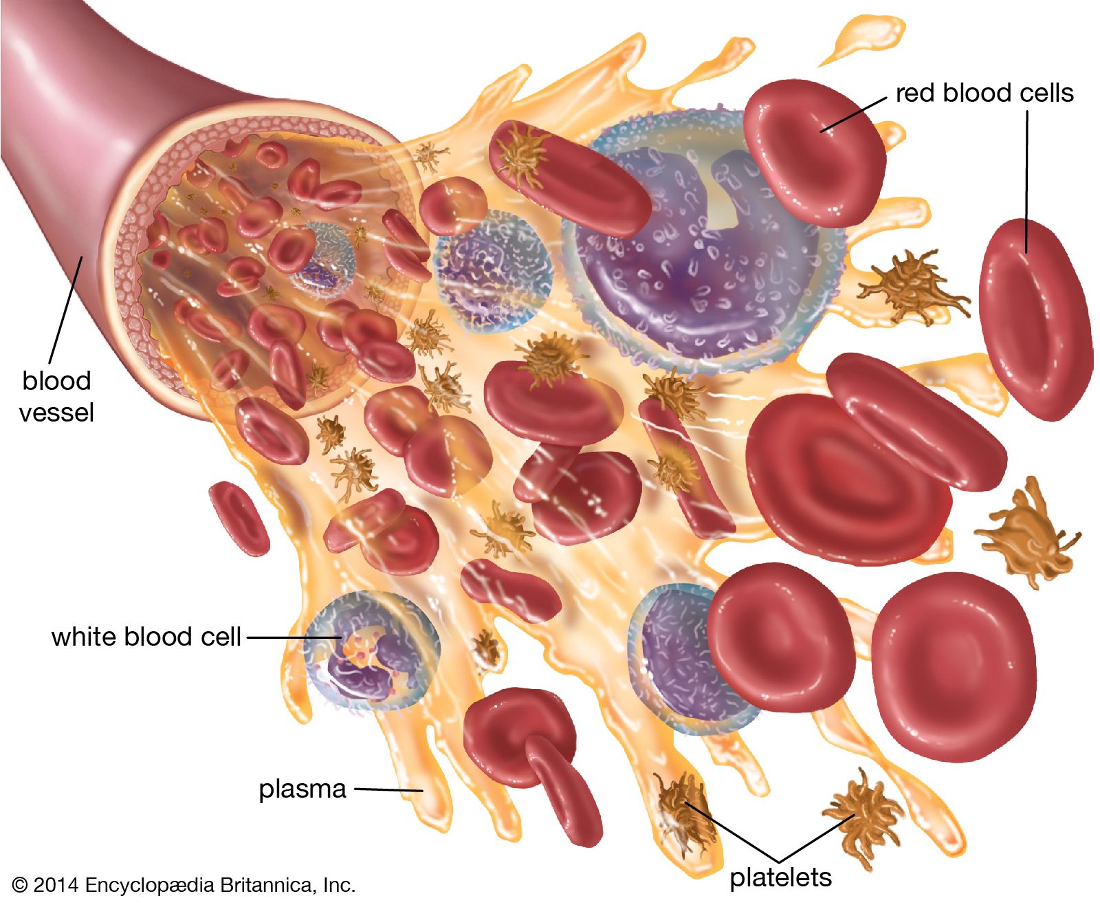


White blood cells (leukocytes)

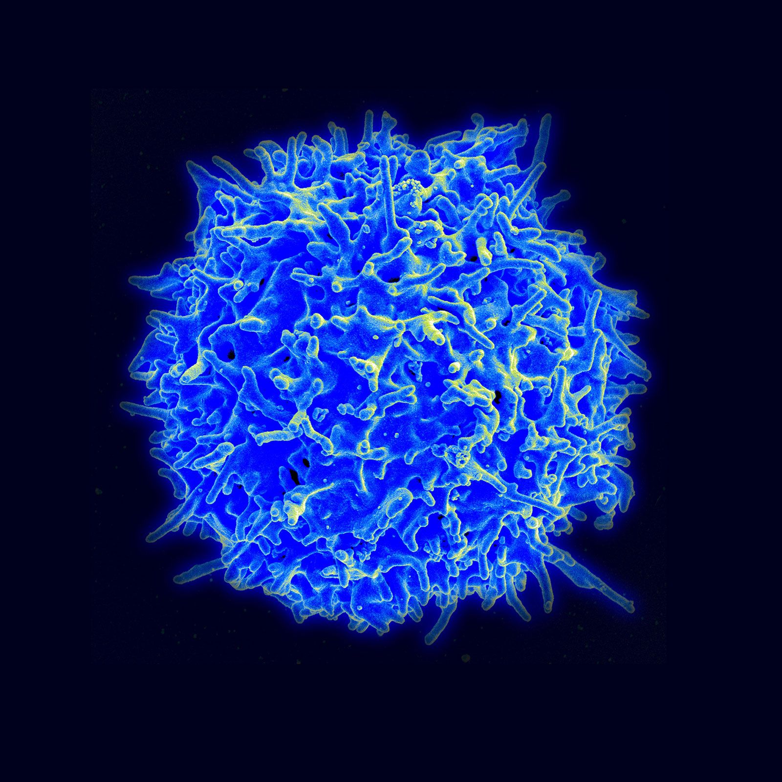
White blood cells (leukocytes), unlike red cells, are nucleated and independently motile. Highly differentiated for their specialized functions, they do not undergo cell division (mitosis) in the bloodstream, but some retain the capability of mitosis. As a group they are involved in the body’s defense mechanisms and reparative activity. The number of white cells in normal blood ranges between 4,500 and 11,000 per cubic millimetre. Fluctuations occur during the day; lower values are obtained during rest and higher values during exercise. Intense physical exertion may cause the count to exceed 20,000 per cubic millimetre. Most of the white cells are outside the circulation, and the few in the bloodstream are in transit from one site to another. As living cells, their survival depends on their continuous production of energy. The chemical pathways utilized are more complex than those of the red cells and are similar to those of other tissue cells. White cells, containing a nucleus and able to produce ribonucleic acid (RNA), can synthesize protein. They comprise three classes of cells, each unique as to structure and function, that are designated granulocytes, monocytes, and lymphocytes.
Granulocytes
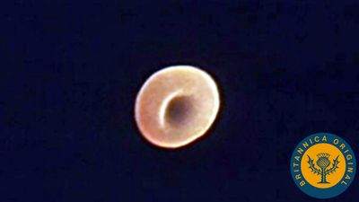
Granulocytes, the most numerous of the white cells, are larger than red cells (approximately 12–15 μm in diameter). They have a multilobed nucleus and contain large numbers of cytoplasmic granules (i.e., granules in the cell substance outside the nucleus). Granulocytes are important mediators of the inflammatory response. There are three types of granulocytes: neutrophils, eosinophils, and basophils. Each type of granulocyte is identified by the colour of the granules when the cells are stained with a compound dye. The granules of the neutrophil are pink, those of the eosinophil are red, and those of the basophil are blue-black. About 50 to 80 percent of the white cells are neutrophils, while the eosinophils and basophils together constitute no more than 3 percent.
Neutrophils
The neutrophils are fairly uniform in size with a diameter between 12 and 15 μm. The nucleus consists of two to five lobes joined together by hairlike filaments. Neutrophils move with amoeboid motion. They extend long projections called pseudopodium into which their granules flow; this action is followed by contraction of filaments based in the cytoplasm, which draws the nucleus and rear of the cell forward. In this way neutrophils rapidly advance along a surface. The bone marrow of a normal adult produces about 100 billion neutrophils daily. It takes about one week to form a mature neutrophil from a precursor cell in the marrow; yet, once in the blood, the mature cells live only a few hours or perhaps a little longer after migrating to the tissues. To guard against rapid depletion of the short-lived neutrophils (for example, during infection), the bone marrow holds a large number of them in reserve to be mobilized in response to inflammation or infection. Within the body, the neutrophils migrate to areas of infection or tissue injury. The force of attraction that determines the direction in which neutrophils will move is known as chemotaxis and is attributed to substances liberated at sites of tissue damage. Of the 100 billion neutrophils circulating outside the bone marrow, half are in the tissues and half are in the blood vessels. Of those in the blood vessels, half are within the mainstream of rapidly circulating blood, and the other half move slowly along the inner walls of the blood vessels (marginal pool), ready to enter tissues on receiving a chemotactic signal from them.
Neutrophils are actively phagocytic; they engulf bacteria and other microorganisms and microscopic particles. The granules of the neutrophil are microscopic packets of potent enzymes capable of digesting many types of cellular materials. When a bacterium is engulfed by a neutrophil, it is encased in a vacuole lined by the invaginated membrane. The granules discharge their contents into the vacuole containing the organism. As this occurs, the granules of the neutrophil are depleted (degranulation). A metabolic process within the granules produces hydrogen peroxide and a highly active form of oxygen (superoxide), which destroy the ingested bacteria. Final digestion of the invading organism is accomplished by enzymes.
Eosinophils
Eosinophils, like other granulocytes, are produced in the bone marrow until they are released into the circulation. Although about the same size as neutrophils, the eosinophil contains larger granules, and the chromatin is generally concentrated in only two nonsegmented lobes. Eosinophils leave the circulation within hours of release from the marrow and migrate into the tissues (usually those of the skin, lung, and respiratory tract) through the lymphatic channels. Like neutrophils, eosinophils respond to chemotactic signals released at the site of cell destruction. They are actively motile and phagocytic. Eosinophils are involved in defense against parasites, and they participate in hypersensitivity and inflammatory reactions, primarily by dampening their destructive effects.
Basophils
Basophils are the least numerous of the granulocytes, and their large granules almost completely obscure the underlying double-lobed nucleus. Within hours of their release from the bone marrow, basophils migrate from the circulation to the barrier tissues (e.g., the skin and mucosa), where they synthesize and store histamine, a natural modulator of the inflammatory response. When aggravated, basophils release, along with histamine and other substances, leukotrienes, which cause bronchoconstriction during anaphylaxis (a hypersensitivity reaction). Basophils incite immediate hypersensitivity reactions in association with platelets, macrophages, and neutrophils.
Monocytes
Monocytes are the largest cells of the blood (averaging 15–18 μm in diameter), and they make up about 7 percent of the leukocytes. The nucleus is relatively big and tends to be indented or folded rather than multilobed. The cytoplasm contains large numbers of fine granules, which often appear to be more numerous near the cell membrane. Monocytes are actively motile and phagocytic. They are capable of ingesting infectious agents as well as red cells and other large particles, but they cannot replace the function of the neutrophils in the removal and destruction of bacteria. Monocytes usually enter areas of inflamed tissue later than the granulocytes. Often they are found at sites of chronic infections.
In the bone marrow, granulocytes and monocytes arise from a common precursor under the influence of the granulocyte-macrophage colony-stimulating factor. Monocytes leave the bone marrow and circulate in the blood. After a period of hours, the monocytes enter the tissues, where they develop into macrophages, the tissue phagocytes that constitute the reticuloendothelial system (or macrophage system). Macrophages occur in almost all tissues of the body. Those in the liver are called Kupffer cells, those in the skin Langerhans cells. Apart from their role as scavengers, macrophages play a key role in immunity by ingesting antigens and processing them so that they can be recognized as foreign substances by lymphocytes.
Lymphocytes

Lymphocytes constitute about 28–42 percent of the white cells of the blood, and they are part of the immune response to foreign substances in the body. Most lymphocytes are small, only slightly larger than erythrocytes, with a nucleus that occupies most of the cell. Some are larger and have more abundant cytoplasm that contains a few granules. Lymphocytes are sluggishly motile, and their paths of migration outside of the bloodstream are different from those of granulocytes and monocytes. Lymphocytes are found in large numbers in the lymph nodes, spleen, thymus, tonsils, and lymphoid tissue of the gastrointestinal tract. They enter the circulation through lymphatic channels that drain principally into the thoracic lymph duct, which has a connection with the venous system. Unlike other blood cells, some lymphocytes may leave and reenter the circulation, surviving for about one year or more. The principal paths of recirculating lymphocytes are through the spleen or lymph nodes. Lymphocytes freely leave the blood to enter lymphoid tissue, passing barriers that prevent the passage of other blood cells. When stimulated by antigen and certain other agents, some lymphocytes are activated and become capable of cell division (mitosis).
The lymphocytes regulate or participate in the acquired immunity to foreign cells and antigens. They are responsible for immunologic reactions to invading organisms, foreign cells such as those of a transplanted organ, and foreign proteins and other antigens not necessarily derived from living cells. The two classes of lymphocytes are not distinguished by the usual microscopic examination but rather by the type of immune response they elicit. The B lymphocytes (or B cells) are involved in what is called humoral immunity. Upon encountering a foreign substance (or antigen), the B lymphocyte differentiates into a plasma cell, which secretes immunoglobulin (antibodies). The second class of lymphocytes, the T lymphocytes (or T cells), are involved in regulating the antibody-forming function of B lymphocytes as well as in directly attacking foreign antigens. T lymphocytes participate in what is called the cell-mediated immune response. T lymphocytes also participate in the rejection of transplanted tissues and in certain types of allergic reactions.
All lymphocytes begin their development in the bone marrow. The B lymphocytes mature partly in the bone marrow until they are released into the circulation. Further differentiation of B lymphocytes occurs in lymphoid tissues (spleen or lymph nodes), most notably on stimulation by a foreign antigen. The precursors of the T lymphocytes migrate from the marrow to the thymus, where they differentiate under the influence of a hormonelike substance. (The thymus is a small organ lying just behind the breastbone in the upper portion of the chest. It is relatively large at birth, begins to regress after puberty, and may be represented only by a fibrous cord in the elderly. The thymus begins to exert its effects on the differentiation of lymphocytes before birth. The removal of the thymus from certain animals at birth prevents the normal development of immunologic responses.) Once they have matured, the T lymphocytes leave the thymus and circulate through the blood to the lymph nodes and the spleen. The two classes of lymphocytes originally derived their names from investigations in birds, in which it was found that differentiation of one class of lymphocyte was influenced by the bursa of Fabricius (an outpouching of the gastrointestinal tract) and thus was called the B lymphocytes, and the other was influenced by the thymus and was called the T lymphocytes.
A primary function of lymphocytes is to protect the body from foreign microbes. This essential task is carried out by both T lymphocytes and B lymphocytes, which often act in concert. The T lymphocytes can recognize and respond only to antigens that appear on cell membranes in association with other molecules called major histocompatibility complex (MHC) antigens. The latter are glycoproteins that present the antigen in a form that can be recognized by T lymphocytes. In effect, T lymphocytes are responsible for continuous surveillance of cell surfaces for the presence of foreign antigens. By contrast, the antibodies produced by B lymphocytes are not confined to recognizing antigens on cell membranes; they can bind to soluble antigens in the blood or in extravascular fluids. T lymphocytes typically recognize antigens of infectious organisms that must penetrate cells in order to multiply, such as viruses. During their intracellular life cycle, viruses produce antigens that appear on the cell membrane. Two classes of T lymphocytes can be involved in the response to those cell-associated viral antigens: cytotoxic T lymphocytes, which destroy the cells by a lytic mechanism, and helper T lymphocytes, which assist B cells to produce antibodies against the microbial antigens. Helper T lymphocytes exert their influence on B lymphocytes through several hormonelike peptides termed interleukins (IL). Five different T lymphocyte interleukins (IL-2, IL-3, IL-4, IL-5, and IL-6) have been discovered, each with different (and sometimes overlapping) effects on B lymphocytes and other blood cells. Interleukin-1, produced by macrophages, is a peptide that stimulates T lymphocytes and that also acts on the hypothalamus in the brain to produce fever. The ability to develop an immune response (i.e., the T cell-mediated and humoral immune responses) to foreign substances is called immunologic competence (immunocompetence). Immunologic competence, which begins to develop during embryonic life, is incomplete at the time of birth but is fully established soon after birth. If an antigen is introduced into a person’s body before immunologic competence has been established, an immune response will not result upon reinfection, and that person is said to be tolerant to that antigen.
Study of immunologic competence and immune tolerance has been accelerated by interest in organ transplantation. The success rates of organ transplantations have been improved by better knowledge about donor selection and improved techniques for suppressing the immune responses of the recipient. An important element in donor selection is tissue typing: the matching of the donor’s histocompatibility antigens (human leukocyte antigens) with those of the prospective recipient. The closer the match, the greater the probability that the graft will be accepted.